|
||
|
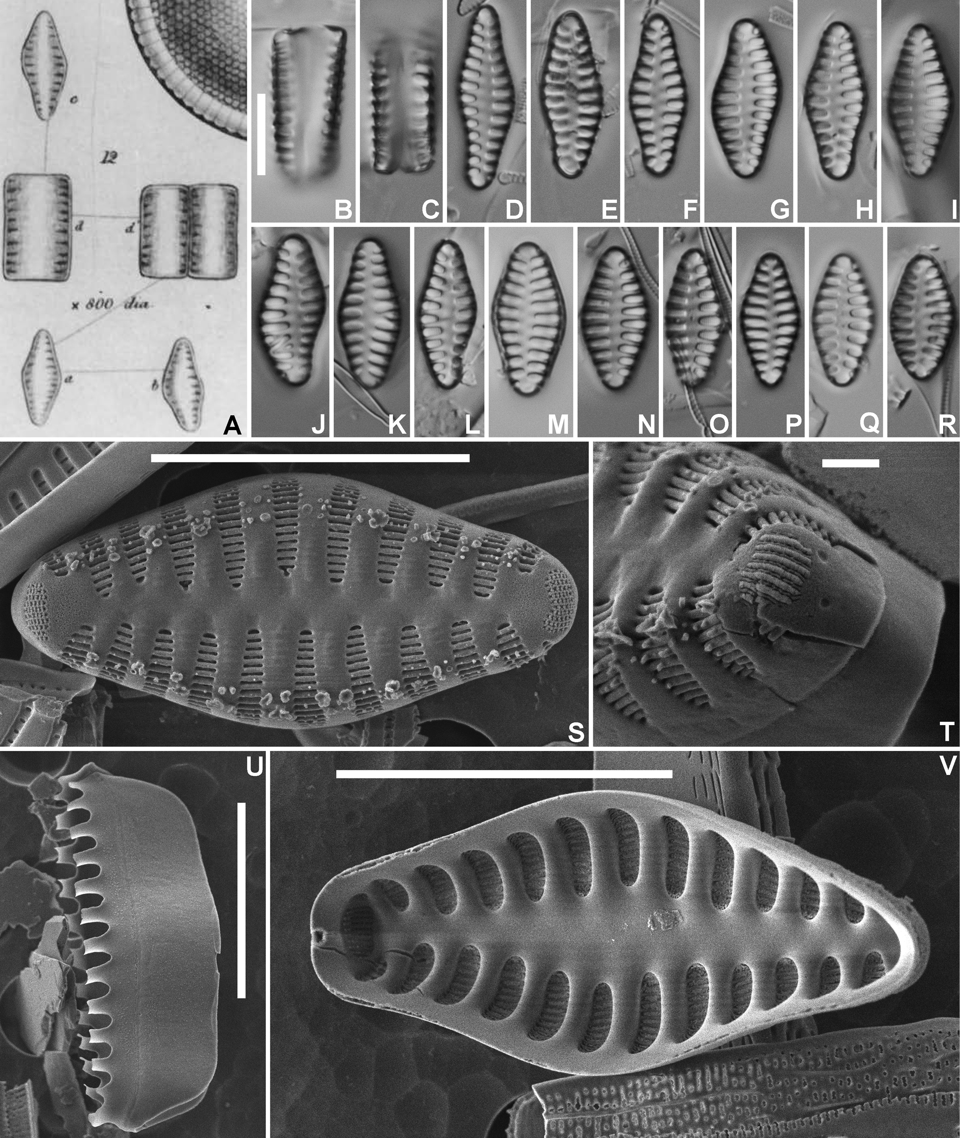
Staurosirella informis (W.Sm.) Van de Vijver comb. nov., LM and SEM micrographs taken from the original Smith material (BR-4821, Cauterets, Gave de Lizez, France). A. Original drawing from Smith (1857: fig. 12). B–C. LM pictures of two frustules in girdle view. D–R. LM pictures of valves in valve face view in decreasing length. S. SEM external view of a complete valve. T. SEM external detail of the footpole showing the large apical pore field. U. SEM view of the valvocopula with the fimbriate extensions. V. SEM internal view of a complete valve. Scale bar = 10 µm (B–S, U–V), 1 µm (T). |