|
||
|
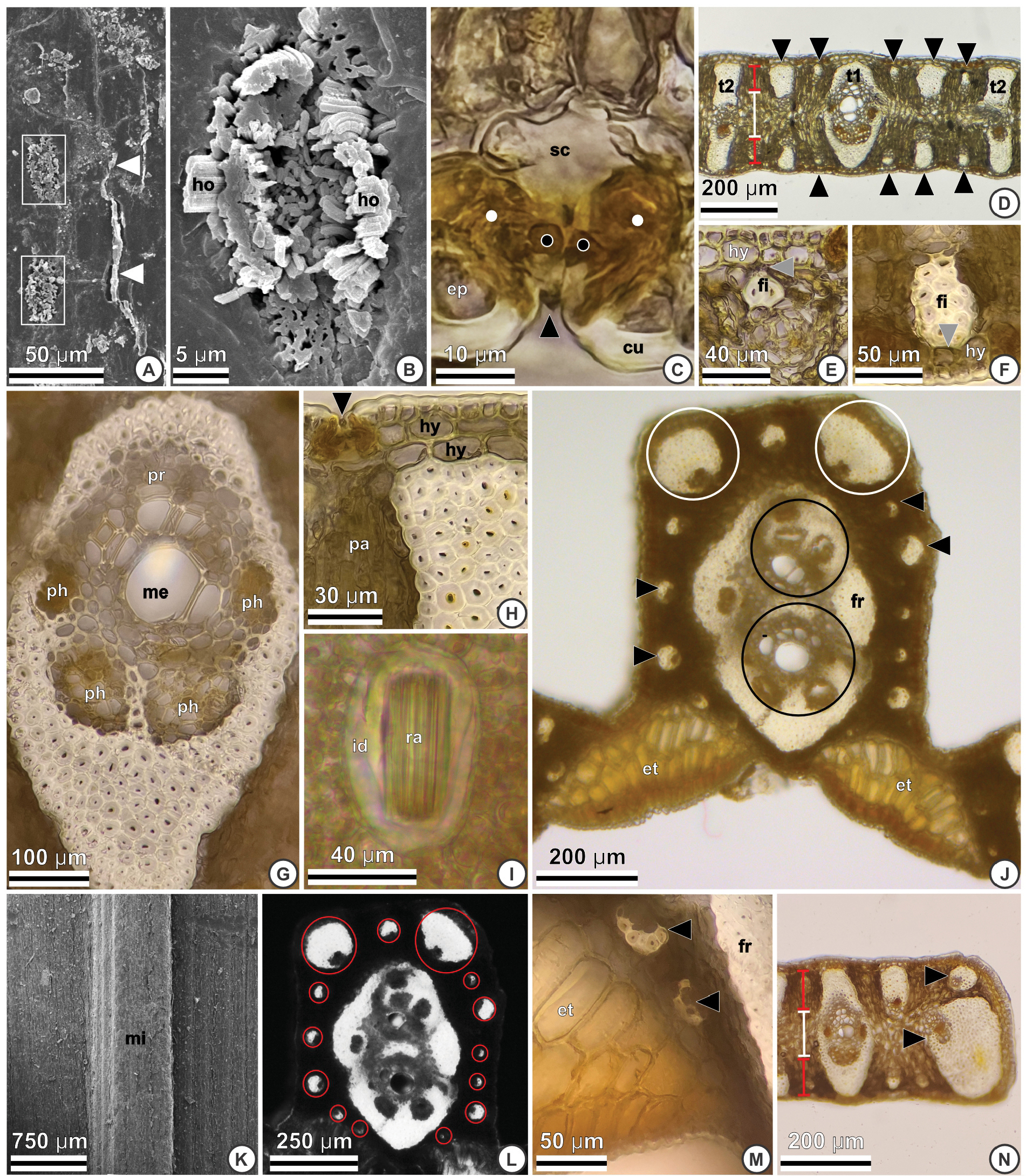
Pinna anatomy of Butia soffiae using SEM (A–B, K) and LM with cross- (C–H, J–N) and longitudinal (I) sections. A. Adaxial surface: horizontal plates of epicuticular waxes (white arrowheads) and stomata (white rectangles). B. Abaxial surface: stomata coated by hook-shaped filaments (ho) of epicuticular waxes. C. Abaxial surface: guard cells (black dots) and lateral subsidiary cells (white dots) are sunken within the epidermis (ep) forming small depressions on the surface (black arrowhead). Cuticle (cu), substomatal chamber (sc). D. Primary (t1), secondary (t2), and tertiary vascular bundles (black arrowheads) connected to the adaxial and abaxial hypodermis; palisade parenchyma (red lines); central parenchyma (white line). E–F. The grey arrowheads indicate the fibres (fi) of the tertiary vascular bundles connected to the adaxial and abaxial hypodermis (hy), respectively. G. Primary vascular bundles with conspicuous metaxylem (me), protoxylem (pr), and four phloem poles (ph). H. Detail of the secondary vascular bundle connected to the adaxial biseriate hypodermis (hy). Stomata (black arrowhead), palisade parenchyma (pa). I. Idioblast (id) containing raphides (ra). J. Expansion tissue (et) interrupted; two collateral bundles (black circles); accessory bundles (black arrowheads), and two accessory bundles with reinforced sheath (white circles); fibrous ring (fr). K. Midrib (mi) adaxially projected, SEM. L. Midrib with all accessory bundles highlighted under polarised light (red circles). M. Detail of expansion tissue cells (et); accessory bundles (black arrowheads) and fibrous ring (fr). N. Pinna margin with reinforced vascular bundles (black arrowheads). Palisade parenchyma (red lines), central parenchyma (white line). |