|
||
|
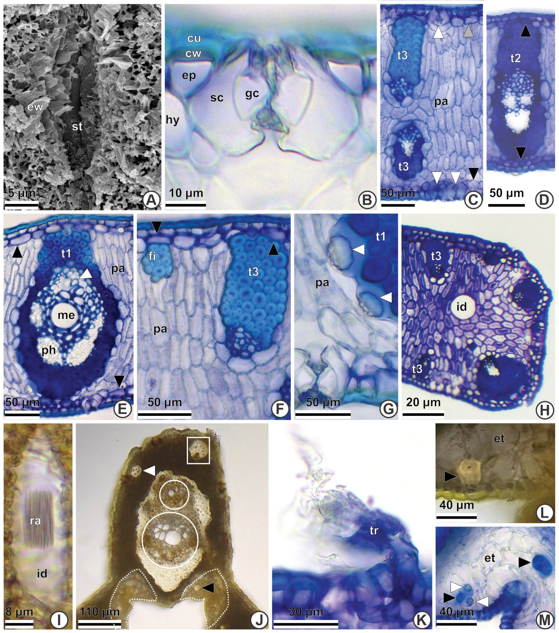
Pinnae anatomy of Syagrus carvalhoi using SEM (A) and LM with cross- (B–H, J–M) and longitudinal (I) sections. A. Adaxial surface: epicuticular waxes (ew) and stomata (st). B. Adaxial surface: guard cells (gc) at the same level of the epidermis (ep); subsidiary cells (sc) at the same level of hypodermis (hy). Cell wall (cw), cuticle (cu). C. Stomata on both surfaces (white arrowheads); palisade parenchyma (pa) throughout the mesophyll; tertiary vascular bundles (t3) connected to the adaxial hypodermis (grey arrowhead) and not connected to the abaxial hypodermis (black arrowhead). D. Secondary vascular bundles (t2) connected to hypodermis (black arrowheads) on both surfaces. E. Primary vascular bundles (t1) connected to the hypodermis (black arrowheads), conspicuous metaxylem (me), protoxylem (white arrowhead) and phloem (ph). F. Adaxial surface: non-vascular fibre bundle (fi) and tertiary vascular bundle (t3) connected to the adaxial hypodermis (black arrowheads). Palisade parenchyma (pa). G. Silica bodies (white arrowheads) associated with primary vascular bundle (t1). Palisade parenchyma (pa). H. Tertiary vascular bundles (t3) and a raphide-containing idioblast (id). I. Idioblast (id) containing raphides (ra). J. Expansion tissue interrupted (surrounded by the dotted white line); fibres in the expansion tissue (white arrowhead); two collateral bundles (white circles); group of fibres (black arrowhead) and accessory bundle (white rectangle). K. Trichome (tr). L. Fibre (black arrowhead) in the expansion tissue (et). M. Silica bodies (white arrowheads) associated with the fibres (black arrowheads) in the expansion tissue (et). |